|
|||||||
Congenital Muscular Torticollis

What is congenital muscular torticollis?
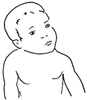
Congenital muscular torticollis or Sternomastoid torticollis is a condition that occurs at birth or up to 2 months of age, where the childís head is tilted to one side. The laymanís term for this condition is wry-neck.
What causes congenital muscular torticollis?
It is traditionally thought to be due to trauma at birth, that causes bleeding in the muscles of the neck, usually the Sternomastoid muscle. The hematoma (blood clot) within the muscle scars down over time, causing the muscle to shorten, thus pulling the head to the typical tilted position. Sometimes, there is an associated mass that can be seen or felt within the Sternomastoid muscle, and usually thought to be a hematoma that is in the process of forming scar tissue. This mass usually disappears by 3 months of age.
More recently, it has been postulated that the sternomastoid muscle shortens as a result of scarring due to an intrauterine vascular disturbance. Still others think that it is due to intrauterine position of the head causing fibrosis or shortening of the muscle.
What are the symptoms?
The condition does not cause pain, but it becomes apparent to the observant mother, as the child persists in holding the head in the tilted position. The right side is involved in 75% of cases, and the child holds his head tilted to the right, with his face and chin rotated to the left. This occurs within the first 2 months of life, and may or may not be associated with a sternomastoid mass. This mass, however, tends to resolve spontaneously after about 3 months.
If uncorrected, as the child grows, the face on the side affected may stay "flattened", so that facial asymmetry is common. This is reversible if the torticollis is corrected before age 1. Beyond that, some facial asymmetry may remain permanent.
What does your doctor do about it?
After a thorough history and examination, your doctor should be able to tell if the torticollis is muscular. There are other causes of torticollis. If the torticollis is due to vertebral abnomalities like atlanto-axial subluxation or hemivertebra, the deformity is quite rigid, and resists any attempt at correcting it passively. Muscular torticollis, even in the more severe cases, can be corrected passively to some extent. In cases where the torticollis is due to spinal cord abnormalities, there are usually other symptoms or signs that suggest the underlying problem. In these cases, X-rays or even MRI exams may be required.
There is a 20% incidence of hip dysplasia in children with muscular torticollis. So it is important that your doctor does an ultrasound exam of the hips in the first 4 to 6 weeks of life to rule that out.
The mainstay of treatment is stretching exercises to stretch the contracted sternomastoid muscle 15 to 20 times, 4 to 6 times a day.
In a child with right torticollis, the head is tilted to the right and the face is rotated to the left. You will want to tilt his head to the left (left ear towards left shoulder), and rotate his face to the right (chin to right shoulder).
In a child with left torticollis, the head is tilted to the left and the face is rotated to the right. You will want to tilt his head to the right (right ear towards right shoulder), and rotate his face to the left (chin to left shoulder).
If you have help with the exercises, your assistant can stabilize the shoulders while you do the stretching exercises with the child lying on his back.
If you do not have help, the exercises can be quite tricky. However, you should still be able to do the exercises adequately by following the instructions for stretching and positioning.
Exercise should be consistent, and generally continued till the child is one year of age. Your doctor or therapist will advise you as to the frequency and duration of the exercises.
What can be expected of the future?
When discovered early, and stretching exercises and positioning followed consistently, 80% recover completely with no long-term effects. In some cases that do not respond to exercises by age 1, surgical release of the sternomastoid muscle may be required.
NOTICE: The information presented is for your information only, and not a substitute for the medical advice of a qualified physician. Neither the author nor the publisher will be responsible for any harm or injury resulting from interpretations of the materials in this article.
Questions
or comments? Post your thoughts in the Orthoseek
Message Forum!
Find a pediatric orthopedic surgeon
in an area near you.
Home | About Us | Orthopaedic Topics | Message Forum
![]()
Comments, questions, or suggestions are welcome. Please
contact us using this form.