|
|||||||
Hip Dysplasia
What is Hip Dysplasia?
Hip Dysplasia is a comprehensive term that has been used to include a spectrum of related developmental hip problems in infants and children, often present at birth. Your doctor may have used one of the following diagnoses for your child instead:
- Congenital hip dislocation - where the hip is frankly dislocated at birth
- Congenital dislocatable hip - where the hip is in place at birth, but dislocates fully when stressed
- Congenital subluxatable hip - where the hip is in place, but dislocates partially when stressed
- Acetabular dysplasia - where the hip socket is shallow and remains shallow, so that the hip is unstable
- Developmental dysplasia (or dislocation) of the hip - a more recent term, to reflect the fact that there are cases that have apparently normal hips at birth, but develop the problem in the first year of life
What causes Hip Dysplasia?
No one knows for sure, but multiple factors are probably involved.
Incidence is 4 per 1000 live-births in the general population, but is much more frequent in Lapps and American Indians. Moreover, the condition tends to run in families and is more common among girls and firstborns. These facts suggest that there is a genetic factor involved.
Certain practices such as infant swaddling and use of the cradle-board in certain cultures increase the chances of developing hip dysplasia. Hence environmental factors are also involved.
Added to these is the observation that during the neonatal period, the baby carries a relatively high level of estrogen from the mother. This relaxes the ligaments In the body. Some babies are especially sensitive to the estrogen, thus causing the hip ligaments to be unduly lax, and the hip "unstable".
What are the symptoms?
The most common scenario is the pediatrician detecting a "hip click" during routine post-natal checkup. Actually, a "clunk", rather than a "click" is detected in the unstable hip, but it requires experience to tell a "clunk" from a "click". The pediatrician typically does the Ortolani test by spreading the thighs, or the Barlow test by bringing the knees together, to elicit this finding.
In the older infant, the pediatrician may suspect the problem if the child has tight hip adductors - he has a hard time spreading the babyís hips. He may also notice that the skin creases around the groin or the buttocks are not symmetrical. Or the legs may appear to be of different lengths.
At age one, the affected child may present with a limp. Unfortunately, pain is not a problem in the child, hence it is easy to miss the diagnosis. However, by the time the patient becomes an adult, arthritic changes will occur, and pain will set in.
How do you prevent it?
It is difficult to prevent something the cause of which is still quite elusive. However, it is well known that in cultures that practice infant swaddling and using cradle boards to carry their babies, the incidence of hip dysplasia is very high. On the other hand, cultures that carry their babies astride the motherís backs have a low incidence of hip dysplasia. Hence it appears logical to discourage putting the babyís legs in the extended position, and encourage keeping the babyís hips spread apart. This latter position places the head of the femur (the ball) against the acetabulum (the socket), and encourages deepening of the socket.
It has been recently recognized that certain babies are more prone to developing hip dysplasia. These "at risk" babies include the following:
- Hip click
- Breech presentation
- Family history of hip dysplasia
- Sternomastoid Torticollis (Wry-neck)
- Foot deformities
- Oligohydramnios (lack of intra-uterine fluid)
A new procedure can now be used as a screening test to check for hip dysplasia in the newborn, using an Ultrasound machine. This is in many ways better than an X-ray examination, which causes radiation and is notorious for being inaccurate for hip dysplasia.
The Ultrasound exam can accurately determine the location of the "ball" in the "socket", the depth of the "socket" and by stressing the hip during the examination, determine the stability of the hip as well. By using sound waves rather than X-rays, there is no risk of radiation to the baby. The Ultrasound exam had now supplanted X-rays in most instances in detecting hip dysplasia in newborns, and is now recommended for all infants "at risk" at around 4 to 6 weeks of age.
What does your doctor do about it?
After taking a history and performing a physical examination, it is likely that he will order an Ultrasound examination of the hips. As mentioned earlier, this has replaced conventional X-rays as a way of confirming the diagnosis. Moreover, it is also a good way of following the progress of the hip with treatment.
The essence of treatment is to reduce the hip in good position, and holding it there by positioning. The position of hip stability is abduction in flexion, and this is the position often used in treatment.
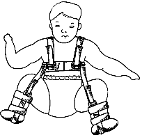
In the first 4 to 6 months of life, the device used is often a Pavlik harness. There have been other devices used in the past, such as the Frejka pillow and the Ilfeld splint, but these are not used as often these days.

The Pavlik Harness was first introduced by Arnold Pavlik, a Czech orthopedic surgeon, who first described it in the 1950ís. Since then, it has become standard treatment for hip dysplasia in infants. The harness consists of a shoulder harness attached to foot stirrups that keep the hips in the position of flexion and abduction, while allowing for a certain degree of movement within the "safe zone". This allows the femoral head (ball) to move within limits of safety within the acetabulum (cup), thus molding and deepening the acetabulum with growth. At birth, the harness is usually worn full-time for 6 weeks when the hip stabilizes, followed by another 6 weeks of weaning. If treatment was started later, or if the hips were more unstable, harness wear might take longer. Your physician will decide on the duration of harness treatment. For details on how to care for your harness, look up The Wheaton-Pavlik Harness: a Guide for Parents.
In the child beyond 6 months, it may not be possible to reduce the hip in a Pavlik harness alone. In these cases, the child may need to be admitted to the hospital and closed reduction performed under general anesthetic. Sometimes a period of leg traction may be needed to facilitate the reduction. Following the reduction, the child is placed in a hip spica cast for about 3 months, followed by the use of a removeable hip abduction brace for another 3 months after that.
In a child older that 1 year, closed reduction alone may not be possible. Open reduction becomes necessary. This involves making an incision to expose the hip at surgery, reducing the hip under direct vision, and stabilizing the hip by reinforcing the hip capsule. The child is then placed in a hip spica cast. In some cases, the hip may redislocate, or continues to stay dysplastic. At that point, further surgery is needed to reconstruct the hip (pelvic or femoral osteotomy or both) to stabilize the hip.
What can be expected after treatment?
For the child discovered to have hip dysplasia within the first 6 weeks of life, treatment in a Pavlik harness is successful in more than 90% of cases. With successful treatment, the hips develop normally, and no long-term problems need be expected.
For the child discovered to have hip dysplasia later in infancy, treatment is more prolonged or complicated, but good results with a normal hip can be expected.
After the age of 1 year, treatment can definitely complicated, and results more guarded. Multiple operations are not unusual, and a normal hip may not result.
The untreated hip causes a limp, which is not painful in childhood. However, arthritis develops early in adult life, and pain sets in. When pain is severe, joint replacement becomes necessary.
NOTICE: The information presented is for your information only, and not a substitute for the medical advice of a qualified physician. Neither the author nor the publisher will be responsible for any harm or injury resulting from interpretations of the materials in this article.
Questions
or comments? Post your thoughts in the Orthoseek
Message Forum!
Find a pediatric orthopedic surgeon
in an area near you.
Home | About Us | Orthopaedic Topics | Message Forum
![]()
Comments, questions, or suggestions are welcome. Please
contact us using this form.